![]()

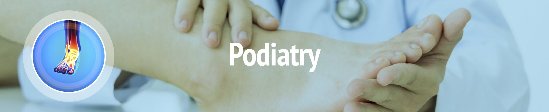
Podiatry is a medical specialty dedicated to diagnosing, treating, and preventing disorders of the foot, ankle, and lower leg. Our podiatrist works with you to manage conditions and infections, offer specialized care for issues like diabetes-related foot complications, and provide orthotics (shoe inserts) to correct biomechanical problems. Our podiatrist provides both general care and surgical treatments for a broad array of foot issues, including skin, nail, and musculoskeletal conditions. They also provide specialized care for chronic diseases that impact the feet.
Depending on the diagnosis, a podiatrist may employ both non-surgical and surgical treatments.
Non-surgical options:
Surgical procedures:
The foot is a complex, flexible structure that contains bones, joints, and more than 100 muscles, tendons and ligaments, all working together to enable movement and balance. The foot is divided into three sections, the forefoot, the midfoot and the hindfoot, which includes the ankle and heel. The heel bone (calcaneus) is the largest bone in the foot.Tendons are bands of tissue that attach muscle to bone. The largest tendon in the body is the Achilles tendon. The Achilles tendon connects the calf muscles to the heel bone, allowing movement such as running, jumping and standing on the toes.
Inflammation is the body’s natural response to injury, disease, overuse or degeneration, and it often causes swelling, pain, or irritation. Inflammation of a tendon is called tendinitis. Achilles tendinitis is a common condition that causes pain along the back of the leg, near the heel. Although the Achilles tendon can withstand great stresses from running and jumping, it is also prone to tendinitis.
Below are some of the most common causes of Achilles tendinitis.
Common symptoms of Achilles tendinitis include:
In most cases, nonsurgical treatment options will provide pain relief, although it may require a few months for symptoms to completely subside. Even with early treatment, the pain of Achilles tendinitis may last longer than 3 months. If appropriate, a foot and ankle conditioning program may be recommended.
Nonsurgical treatment may include:
Surgical treatment to relieve Achilles tendinitis should only be considered if pain does not improve after 6 months of nonsurgical treatment.
With any surgery there are some risks, and these vary from person to person. Complications are typically minor, treatable and unlikely to affect your final outcome. Your orthopaedic surgeon will speak to you prior to surgery to explain any potential risks and complications that may be associated with your procedure.
For additional information, we have included this patient education sheet as a pdf to view, download and print:
The ankle joint connects the leg and the foot. It is formed by three separate bones, the tibia, fibula and talus. The shinbone (tibia) supports most of a person’s weight when standing. The outer bone (fibula) is the smaller bone of the lower leg. A small, irregular-shaped foot bone (talus) connects the tibia and fibula. Acting as a hinge, these bones form the ankle. The ankle joint allows movement such as walking, running and jumping, and also contributes to lower limb stability.The ankle is reinforced by fibrous tissue (ligaments) that connects bone to bone. Ligaments have an elastic structure that allows them to stretch, within their limits, and then return to their normal positions. Ligaments protect the ankle from abnormal movements-especially twisting, turning and rolling of the foot.
Arthritis is inflammation that can cause pain and stiffness in any joint in the body. Osteoarthritis, also known as degenerative or “wear-and-tear” arthritis, is a common problem for many people after reaching middle age. It is often experienced in the small joints of the foot and ankle. In osteoarthritis, the cartilage in the joint gradually wears away, becoming frayed and rough. As protective space between the bones decreases, it can result in bone rubbing on bone, causing painful osteophytes (bone spurs).
Your physician may suggest changes in your daily lifestyle that can help relieve the pain of arthritis and slow the progression of the disease. If appropriate, a foot and ankle conditioning program may be recommended.
If your pain causes disability and is not relieved by nonsurgical treatment, surgery may be recommended. The appropriate surgery will depend on the type and location of the arthritis and the impact of the disease on your joints. In some cases, more than one type of surgery may be recommended.
With any surgery there are some risks, and these vary from person to person. Complications are typically minor, treatable and unlikely to affect your final outcome. Your orthopaedic surgeon will speak to you prior to surgery to explain any potential risks and complications that may be associated with your procedure.
The ankle joint connects the leg and the foot. It allows movement such as walking, running and jumping, and also contributes to lower limb stability. The ankle joint is formed by three separate bones, the tibia, fibula and talus. The shinbone (tibia) supports most of a person’s weight when standing. The outer bone (fibula) is the smaller bone of the lower leg. A small, irregular-shaped foot bone (talus) connects the tibia and fibula. Acting as a hinge, these bones form the ankle.
The ankle is reinforced by fibrous tissue (ligaments) that connects bone to bone. Ligaments have an elastic structure that allows them to stretch, within their limits, and then return to their normal positions. Ligaments protect the ankle from abnormal movements—especially twisting, turning and rolling of the foot.
When a ligament is forced to stretch beyond its normal range, a sprain occurs. A severe sprain causes actual tearing of the elastic fibers of the ligament. A sprained ankle is a very common injury that produces pain and swelling. If the sprain is a result of excess force, you may hear a “pop” sound when the injury occurs. The grade, or severity, of the sprain is determined by the amount of force that caused the injury.
The amount of pain and tenderness resulting from a strain depends on the amount of stretching and tearing of the ligament. The ankle may be swollen and painful, and walking may be difficult. Instability occurs when there has been complete tearing of the ligament or a complete dislocation of the ankle joint.
A sprained ankle can happen to anyone, child or adult, athlete or not. It can occur during sports and physical fitness activities, or it can be the result of something as simple as stepping on an uneven surface or stepping down at an angle. When the foot twists, rolls or turns beyond its normal range of motion and the ligaments stretch in an extreme or abnormal position, the ankle may be sprained.
If an ankle sprain is not recognized and treated with the necessary attention and care, chronic problems of pain and instability may result, so it is important to seek care right away. A broken bone or fracture can have similar symptoms of pain and swelling, so your physician may order X-rays to be sure there are no broken bones in the ankle or foot. Once a break can be ruled out, your physician may be able to diagnose the grade of the sprain based on the amount of swelling, pain and bruising. Although the ankle may be tender or painful, it may be necessary to move it in various ways during the exam to determine which ligament has been hurt or torn. After swelling and bruising subsides, an MRI (magnetic resonance imaging) scan may be needed to help ensure a correct diagnosis if your physician suspects a severe injury to the ligaments, injury to the joint surface, a small bone chip, or other problems.
Most ankle sprains need only a period of protection to heal. Swelling and pain usually last 2 to 3 days, however, the healing process takes about 4 to 6 weeks. During this time, use rest, ice, compression and elevation (R.I.C.E.) to help with pain and swelling, and you may also need to use crutches if walking causes pain.
A Grade 1 sprain is commonly treated with R.I.C.E. If your sprain is Grade 2, it make take longer for healing to occur and your physician may use a splint or other device to immobilize the ankle. Grade 3 sprains can be associated with permanent instability. Surgery is rarely needed, however a short leg cast or cast-brace may be used for 2 to 3 weeks.
Surgery is reserved for injuries that fail to respond to nonsurgical treatment, and for persistent instability following months of rehabilitation and nonsurgical treatment. Surgical options include:
Arthroscopy—During this minimally invasive surgical procedure, the surgeon looks inside the joint to see if there are any loose fragments of bone or cartilage, or part of the ligament caught in the joint.
Reconstruction—A surgeon repairs the torn ligament with stitches or sutures, or uses other ligaments and/or tendons found in the foot and around the ankle to repair the damaged ligaments.
With any surgery there are some risks, and these vary from person to person. Complications are typically minor, treatable and unlikely to affect your final outcome. Your orthopaedic surgeon will speak to you prior to surgery to explain any potential risks and complications that may be associated with your procedure.
The length of time you can expect to spend recovering after surgery will depend on the extent of injury and the amount of surgery that was required. Rehabilitation to restore strength and range of motion to a level that allows you to return to pre-injury function may take from weeks to months. If appropriate, a foot and ankle conditioning program may also be prescribed.
The best way to prevent ankle sprains is to maintain good strength, muscle balance and flexibility, as follows:
Nearly one-fourth of all bones in the human body are in the feet. The foot is a complex, flexible structure that contains bones, joints, and more than 100 muscles, tendons and ligaments, all working together to enable movement and balance. The foot is divided into three sections, the forefoot, midfoot and hindfoot. The forefoot has five toes (14 phalanges) and five longer bones (metatarsals). One phalanx of each of the five toes connects to one of the five metatarsals.
The big toe, or great toe (hallux), is made up of two joints. The metatarsophalangeal joint (MTP) is the largest of these, and the closest to the base of the toe, where the first long bone of the foot (metatarsal) meets the first bone of the toe (phalanx). In the MTP joint, as in any joint, the ends of the bones, where they touch, are covered by articular cartilage, a smooth substance that protects the bones and enables them to move easily.
A bunion is a bump on the MTP joint, on the inner border of the foot. Bunions are made of bone and soft tissue, covered by skin that may be red and tender.
A bunion may be sore and swollen. If you have a bunion, it may hurt to wear any type of shoe. The MTP joint flexes with each step, so as the bunion becomes larger, walking becomes increasingly painful and bursitis may also set in. A bunion may cause the big toe to angle toward the second toe, or possibly move all the way under it. Pressure from the big toe may force the second toe out of alignment, sometimes causing it to overlap the third toe. Skin on the bottom of the foot may thicken and become painful. An advanced bunion can make the foot look grotesque and if it becomes too severe, walking may be difficult. Pain may become chronic and you may develop arthritis.
Prevention is best, so minimize your chances of developing a bunion by never forcing your feet into shoes that fit improperly, are short, tight, sharply pointed, or have heels higher than 2 1/4 inches. Instead, choose shoes with wide insteps, broad toes and soft soles that conform to the natural shape of the feet.
If you already have a bunion and it is not too severe, it may be treatable without surgery. Wearing shoes that are roomy enough to not put pressure on the bunion should help relieve pain. Protective pads can be used to cushion the painful area, and you may also want to consider having your shoes stretched professionally. If the bunion causes difficulty walking or produces pain despite wearing accommodating shoes, you may need surgery.
Orthopaedic surgeons use various surgical procedures to treat bunions, but the common goal for them all is to realign the joint, relieve pain, and correct deformity. Your orthopaedic surgeon will choose the procedure that is best suited to your condition.
With any surgery there are some risks, and these vary from person to person. Complications are typically minor, treatable and unlikely to affect your final outcome. Your orthopaedic surgeon will speak to you prior to surgery to explain any potential risks and complications that may be associated with your procedure.
For additional information, we have included this patient education sheet as a pdf to view, download and print:
A callus is a hard pad of thickened skin that develops as the skin tries to protect itself from repeated friction, pressure, rubbing or irritation. Calluses are most often found on pressure spots, including the heels, balls and sides of the feet, as well as the big toe. Calluses are larger than corns, vary in size and shape, and are not typically painful. Some callus formation on the sole of the foot is normal and may disappear once the source of irritation is eliminated.
Corns are smaller and deeper than calluses and often form on the top or side of a toe. They have a hard center surrounded by swollen skin and are painful when pressed.
There are different types of corns:
Poorly fitting shoes are the most common cause of calluses and corns, but they can also result from abnormal foot function or high levels of repetitive activity.
Causes and risk factors include:
While both can be painful, calluses are typically less sensitive than corns. Untreated, both calluses and corns may become more painful as they thicken over time.
In many cases, corns and calluses disappear once the source of friction has been eliminated. If you have diabetes or poor blood flow, it is extremely important to see your physician immediately, before attempting to treat the condition yourself.
If the corn or callus becomes painful or inflamed, or self-care with a pumice stone and soaking is unsuccessful, medical treatment may be required for relief, such as:
If you have a structural deformity of the foot or toes that is causing development of corns or calluses, your specialist may recommend surgery to realign or remote bone tissue. Surgery may also be recommended if corns or calluses are causing extreme pain, preventing a normal gait or comfortable walking, or resulting in infections or other issues.
Nearly one-fourth of all bones in the human body are in the feet. The foot is a complex, flexible structure that contains bones, joints, and more than 100 muscles, tendons and ligaments, all working together to enable movement and balance. The foot is divided into three sections:
Difficulties with foot position and function can lead to more serious problems, not only for the feet, but also for other areas, including the spine. In some cases, these problems may be caused by footwear that fits improperly, does not accommodate normal foot alignment, or that interferes with natural movement and balance of the body.
The term “cavovarus” refers to a foot with an arch that is higher than normal, and that turns in at the heel. This is a deformity that tends to worsen gradually over time. Depending on the age of the patient and the degree of deformity and stiffness, treatment options may range from supportive care with bracing, to surgical treatment for soft tissue releases, tendon transfers, and possibly reshaping or fusion of the bones and joints.
As the deformity worsens, there can be increasing pain at the ankle due to recurrent sprains, painful calluses at the side of the foot or base of the toes, or difficulty with shoe wear.
Weakness in the peroneal muscles and sometimes the small muscles in the foot are often the cause of a cavovarus foot deformity.
Initially, a careful investigation is needed to rule out any neurological condition that may be causing the foot’s high arch. Your doctor will examine your foot and gait, observing as you walk and stand to determine the shape of the arch and heel position. Mobility of the heel will be checked with an exam called The Coleman Block Test, and X-rays may be needed to allow your physician to review the shape of the bones. Often, the bones and joints appear normal other than alignment with the high arch and inward rotation of the heel.
Treatment will depend on what, if anything, is causing pain. Generally, treatment of the foot deformity can involve several options. In mild cases, foot pain can be addressed with orthotics or custom shoes to support and protect the foot and relieve pressure areas. Corns and calluses, if present, can be treated with a regular skin care routine. If appropriate, a foot and ankle conditioning program may be recommended.
In severe cases, especially if pain is present and the height of the arch is progressively increasing, surgical treatment may be recommended. This can involve release of contracted soft tissues, tendon transfers to rebalance the foot, osteotomies to reshape the foot, and possibly joint fusions to realign and hold the foot in a corrected position.
With any surgery there are some risks, and these vary from person to person. Complications are typically minor, treatable and unlikely to affect your final outcome. Your orthopaedic surgeon will speak to you prior to surgery to explain any potential risks and complications that may be associated with your procedure.
Nearly one-fourth of all bones in the human body are in the feet. The foot is a complex, flexible structure that contains bones, joints, and more than 100 muscles, tendons and ligaments, all working together to enable movement and balance. The foot is divided into three sections, the forefoot, midfoot and hindfoot. The forefoot has five toes (14 phalanges) and five longer bones (metatarsals). One phalanx of each of the five toes connects to one of the five metatarsals.
Also referred to as Charcot arthropathy or Charcot neuropathy, Charcot foot is a rare, but very serious complication of diabetic neuropathy, which causes nerve damage that results in a loss of feeling in the lower legs, ankles and feet. This makes it difficult, or impossible, to feel pain, temperature, or symptoms of other problems with your feet. Unnoticed and untreated, even a slight injury can become infected and continue to worsen. Charcot foot occurs when an injury or infection causes serious complications, such as weakened bones, fractures, collapsed joints, deformities of the foot, or infection that spreads to the rest of the body. Severe cases of Charcot foot can lead to disability, life-threatening problems, or amputation of the foot. Those who have had neuropathy for a long period of time are at higher risk for developing Charcot foot, especially those with a tight Achilles tendon.
The food (mainly carbohydrates) we eat provides glucose, which the body’s cells require for energy. Insulin, a hormone produced by the pancreas gland, helps glucose to enter the cells from the bloodstream. When blood glucose levels rise, such as after a meal, the pancreas releases insulin, which helps lower the level of glucose in the blood by allowing it to enter cells. Without insulin, glucose builds up in the blood. Diabetes occurs when blood glucose, or blood sugar, levels are too high (hyperglycemia). This disease can cause nerve damage, poor circulation, and a weakened immune system, making it hard for the body to deliver oxygen and nutrients to a wound or injury. Diabetes can slow the healing process and make it difficult for the body to fight infections.
Immediate treatment is essential in preventing complications, so it is crucial to see your podiatrist if any symptoms appear, in addition to your regular check-ups.
Early symptoms include:
Symptoms of more advanced Charcot foot include:
Charcot foot most often affects people with diabetes, especially those who have had it for seven years or more or have uncontrolled or difficult-to-manage blood sugar levels. Smoking and regular use of alcohol increases the risk.
In addition to diabetes, other health conditions increase the risk of developing Charcot foot, including:
Prevention of severe complications such as loss of a toe, foot, or leg is the primary objective of Charcot foot treatment. Your physician will recommend appropriate treatments to provide relief of symptoms and help reduce permanent issues that impair use of the foot.
Common treatments include:
If your Charcot foot is advanced and you’re experiencing severe symptoms, such as serious infection, collapsed bones and joints, or ulcers, surgery may be required to stabilize the foot and repair damage within the foot and ankle. Although rare, in the most severe and advanced cases, amputation may be necessary. Your surgeon will discuss any recommended procedures with you, including the benefits, potential risks, and what you can expect during recovery.
The best way to prevent Charcot foot is to manage your blood sugar (blood glucose) levels, have regular checkups with your podiatrist, and examine your feet for any loss or change in your ability to feel touch, pain or pressure.
Prevention and proper foot care includes:
Complex regional pain syndrome (CRPS) is an uncommon, physiological condition that causes chronic pain, most often in the arms, legs, hands or feet, but it can occur anywhere in the body. Although CRPS typically follows an injury or surgery, it can also develop after a minor injury, and in rare instances, without apparent cause. CRPS can be an acute, or short-term condition, or it can be chronic, lasting three months or more. The pain of CRPS is usually much greater than that of the initial trauma. Symptoms usually improve over time, but many still experience some degree of pain for a year or more.
CRPS Type 1—Also referred to as sympathetic dystrophy; develops without known nerve damage
CRPS Type 2—Also referred to as causalgia; follows damage to a specific nerve
The symptoms and stages for both types of CRPS are the same, however triggers vary, and the pain can be continuous, or it can come and go. Most people do not experience all the symptoms listed below. CRPS typically follows three stages of development.
Stage I: Acute—Usually lasts from 1-3 months
Stage II: Dystrophic—Usually lasts from 3-12 months
Stage III: Atrophic—Occurs after 1 year
The exact cause of CRPS is unknown, however, both types can typically be linked to illness, injury or trauma, such as: fractures, sprains, strains, surgery, being in a cast, nerve injuries or pressure on a nerve, burns, cuts, infection, stroke, heart attack, cancer, or neck problems.
The risk of CRPS also increases for those who smoke or have autoimmune disorders, diabetes, poor circulation, or previous nerve damage. If a sibling or other close family member has CRPS, you may have a higher risk for developing the condition at an early age, however, genetic aspects of CRPS are still being studied.
If you’re experiencing severe, continuous pain made worse by touching or movement of the affected area, see your healthcare provider to determine the cause. Early diagnosis and treatment is crucial to limiting the progression of CRPS and preventing additional, and sometimes disabling, symptoms of this condition.
Your physician may conduct a physical examination based on a set of guidelines known as the Budapest Criteria. Developed by the International Association for the Study of Pain (IASP), the Budapest Criteria differentiates between signs (seen or felt by the examining medical professional) and symptoms (reported by the patient to the physician), and provides a framework for accurate diagnosis of CRPS.
Currently, CRPS cannot be definitively diagnosed using a single test, however, specific tests may be needed to rule out other possible causes of pain and symptoms. Before a diagnosis of CRPS is made, other causes of the signs and symptoms presented are eliminated. This may require blood tests to rule out rheumatoid arthritis or an infection. Diagnostic imaging studies may also be needed, such as X-rays, magnetic resonance imaging (MRI), or bone density scans, as well as other types of testing.
For some people, the symptoms of CRPS go away on their own, however, there is no cure at present. Others may experience persistent symptoms for months, or even years. Treatment is typically most effective when started early in the course of the illness.
Rehabilitation or occupational therapy—Your physical medicine and rehabilitation (PM&R) specialist may recommend special exercises to help you maintain function, flexibility and strength, improve blood flow, and prevent loss of muscle tissue in the affected limb. Specifically, desensitization therapy has shown to provide a significant benefit in the treatment of CRPS.
Medications—Over-the-counter, nonsteroidal anti-inflammatories (NSAIDs) such as ibuprofen and naproxen may be prescribed alone to treat moderate pain and inflammation, or in combination with acetaminophen, lidocaine and other topical pain relievers, and other treatments, such as corticosteroids. If you have serious contraindications to NSAIDs, other types of pain medication can be considered.
Sympathetic Nerve Blocks—Sympathetic nerve fibers converge at specific areas in the body, known as a ganglion or plexus. In the setting of CRPS symptoms that do not respond to conservative treatment, the ganglion/plexus can be targeted with a fluoroscopic (X-ray) guided injection. Examples of the targeted area include the stellate ganglion, celiac plexus, lumbar sympathetic plexus, and the superior hypogastric plexus. The injection typically consists of local anesthetic and can be a helpful tool for both diagnosis and treatment.
Peripheral Nerve Stimulation (PNS)—For patients with persistent symptoms that are not responsive to conservative treatment, do not require surgery, and have a known nerve injury causing their CRPS, peripheral nerve stimulation can be a viable option. This consists of your doctor implanting an electrode beneath your skin next to the damaged nerve. Electrical stimulation is then utilized to disrupt the pain signals coming from the damaged nerve for improved pain and function. Depending on the device, some require an external battery that is worn over the damaged nerve or implanted beneath the skin.
Dorsal Column Spinal Cord Stimulation (SCS)—Spinal cord stimulation (SCS) has been a long-standing, FDA approved treatment for upper and lower extremity CRPS. If patients have persistent symptoms despite extensive conservative treatment, they may be a candidate SCS. In a two-step process, your doctor will typically implant 2 electrodes underneath the skin in an area behind the spinal cord known as the epidural space. These will remain in place for up to 1 week with an external battery that is worn on top of the skin. The goal is to disrupt the pain signals and provide improved pain and function. If the patient achieves at least 50% relief in their symptoms during the trial, they would be a candidate for permanent implantation of the leads and battery underneath the skin.
Dorsal Root Ganglion (DRG) Stimulation—In cases where there is a peripheral nerve injury contributing to the CRPS symptoms, certain peripheral nerves are difficult to access for peripheral nerve stimulation. In such cases, stimulation can be achieved by targeting the nerve fibers as the exit out of the spinal canal, known as the dorsal root ganglion (DRG). DRG stimulation can also be used in situations where there is no direct nerve injury (CRPS type 1). In a two-step process, your doctor will typically implant 2 electrodes underneath the skin next to the target DRG. These will remain in place for up to 1 week with an external battery that is worn on top of the skin. The goal is to disrupt the pain signals and provide improved pain and function. If the patient achieves at least 50% relief in their symptoms during the trial, they would be a candidate for permanent implantation of the leads and battery underneath the skin. This is FDA approved for lower extremity CRPS only.
Nearly one-fourth of all bones in the human body are in the feet. These complex, flexible structures contain bones, joints, and more than 100 muscles, tendons and ligaments, all working together to enable movement and balance. Difficulties with the feet and toes can lead to more serious problems, not only for the feet, but also for other areas of the body, including the hips and spine.
The food (mainly carbohydrates) we eat provides glucose, which the body’s cells require for energy. Insulin, a hormone produced by the pancreas gland, helps glucose to enter the cells from the bloodstream. When blood glucose levels rise, such as after a meal, the pancreas releases insulin, which helps lower the level of glucose in the blood by allowing it to enter cells. Without insulin, glucose builds up in the blood. Diabetes occurs when blood glucose, or blood sugar, levels are too high. This disease can cause nerve damage, poor circulation, and a weakened immune system, making it hard for the body to deliver oxygen and nutrients to a wound or injury. Diabetes can slow the healing process and make it difficult for the body to fight infections.
Diabetic foot is a condition that can cause foot ulcers. It refers to a variety of foot complications caused by the damage to nerves and blood vessels that occurs over time from high blood sugar levels. Diabetic foot can cause pain, numbness, tingling, and loss of feeling in the feet and without normal pain sensation, a cut, blister, sore, or ulcer on the foot may go unnoticed.
Because diabetes slows the healing process, a sore or ulcer that doesn’t receive proper wound care can worsen and become infected, leading to serious complications, including gangrene and amputation.
If you have diabetes, you are at risk for diabetic foot, peripheral neuropathy, Charcot foot, or nerve damage. However, there are additional factors that further increase your risk of developing diabetic foot, such as uncontrolled or difficult-to-manage blood sugar levels and number of years living with diabetes.
Knowing what issues to look for, how to care of your feet properly, and when to see prompt treatment will help you avoid serious problems.
Examine your feet daily and see your physician immediately if you experience any of the following:
Healing a diabetic foot ulcer as quickly as possible lessens the chance of infection and serious complications.
Treatment may include:
Proper Foot Care and Preventing Diabetic Foot
The food (mainly carbohydrates) we eat provides glucose, which the body’s cells require for energy. Insulin, a hormone produced by the pancreas gland, helps glucose to enter the cells from the bloodstream. When blood glucose levels rise, such as after a meal, the pancreas releases insulin, which helps lower the level of glucose in the blood by allowing it to enter cells. Without insulin, glucose builds up in the blood. Diabetes occurs when blood glucose, or blood sugar, levels are too high (hyperglycemia). This disease can cause nerve damage, poor circulation, and a weakened immune system, making it hard for the body to deliver oxygen and nutrients to a wound or injury. Diabetes can slow the healing process and make it difficult for the body to fight infections.
Over time, high blood sugar levels associated with diabetes can damage the nerves throughout the body, resulting in a serious complication known as diabetic neuropathy. Diabetic peripheral neuropathy is the most common type of neuropathy seen by podiatrists because it causes nerve damage in the feet and lower limbs.
Knowing what issues to look for and how to care of your feet properly will help you avoid serious problems.
Some of the most commonly experienced symptoms include:
If you have diabetes, you are at risk for diabetic neuropathy or nerve damage. However, there are additional factors that further increase your risk, such as uncontrolled or difficult-to-manage blood sugar levels and number of years living with diabetes. Additional risk factors include high blood pressure, high cholesterol levels, obesity, and smoking.
Diabetic neuropathy can range from mild to severe but tends to worsen if steps aren’t taken to treat it. Your podiatrist can help you manage diabetic neuropathy by monitoring the health of your feet, prescribing orthotics, taking care of any injuries or foot wounds, and much more. Although there is no cure for diabetic neuropathy, proper treatment can address symptoms and slow progression.
If you’re in the early stages of the condition, conservative measures may be recommended, such as:
For advanced, more severe, or cases unresponsive to nonsurgical treatments, surgery may be recommended to correct any foot deformities or remove infected tissue. Your podiatrist will discuss the potential advantages and risks of any surgery with you.
Proper Foot Care and Preventing Complications
The ankle joint connects the leg and the foot. It is formed by three separate bones, the tibia, fibula and talus. The shinbone (tibia) supports most of a person’s weight when standing. The outer bone (fibula) is the smaller bone of the lower leg. A small, irregular-shaped foot bone (talus) connects the tibia and fibula. Acting as a hinge, these bones form the ankle. The ankle joint allows movement such as walking, running and jumping, and also contributes to lower limb stability.
The ankle is reinforced by fibrous tissue (ligaments) that connects bone to bone. Ligaments have an elastic structure that allows them to stretch, within their limits, and then return to their normal positions. Ligaments protect the ankle from abnormal movements—especially twisting, turning and rolling of the foot.
When the ankle joint lacks flexibility and upward, toes-to-shin movement of the foot (dorsiflexion) is limited, the condition is called equinus. Equinus is a result of tightness in the Achilles tendon or calf muscles (the soleus muscle and/or gastrocnemius muscle) and it may be either congenital or acquired. This condition is found equally in men and women, and it can occur in one foot, or both. If both feet are involved, range of motion may be more limited in one foot than in the other. This limited muscle flexibility and range of motion can lead to injury.
People with equinus develop ways to compensate for their limited ankle motion, which often leads to other problems of the foot, leg or back. Common methods of compensation include: flattening the arch, picking up the heel early when walking, and putting increased pressure on the ball of the foot. In addition to contributing to a number of foot and ankle problems, equinus can hinder the healing of a foot or ankle injury.
Depending on how a patient compensates for the inability to bend properly at the ankle, a variety of other foot conditions can develop, such as:
A person with equinus has a limited range of ankle motion and lacks the flexibility needed to bring the top of the foot upward, toward the shin (tibia).
Equinus is often it is due to tightness in the Achilles tendon or calf muscles. For some, this may be congenital (present at birth) or an inherited trait. For others, this tightness is acquired and the result of being in a cast or on crutches, or frequently wearing high-heeled shoes. Other causes include diabetes or having one leg shorter than the other. If a bone or bone fragment (following an ankle injury, for example) blocks movement of the ankle, the patient may experience equinus. Infrequently, equinus can be caused by spasms in the calf muscle, which may be a sign of an underlying neurologic disorder.
Most patients with equinus are diagnosed when seeking treatment for other foot problems associated with equinus. During the examination, your orthopaedic surgeon will evaluate the ankle’s range of motion when the knee is flexed (bent), and when it is extended (straightened). This enables the surgeon to identify whether the tendon or muscle is tight, and to assess whether bone is interfering with ankle motion. X-rays may also be ordered. In some cases, the foot and ankle surgeon may refer the patient for neurologic evaluation.
Some nonsurgical treatment strategies are aimed at relieving the symptoms and conditions associated with equinus. Treatment for the equinus itself may include one or more of the following options.
In some cases, surgery may be needed to correct the cause of equinus if it is related to a tight tendon or a bone blocking the ankle motion. Your orthopaedic surgeon will determine the type of procedure that is best suited for you.
With any surgery there are some risks, and these vary from person to person. Complications are typically minor, treatable and unlikely to affect your final outcome. Your orthopaedic surgeon will speak to you prior to surgery to explain any potential risks and complications that may be associated with your procedure.
Nearly one-fourth of all bones in the human body are in the feet. The foot is a complex, flexible structure that contains bones, joints, and more than 100 muscles, tendons and ligaments, all working together to enable movement and balance. The foot is divided into three sections, the forefoot, midfoot and hindfoot. The forefoot has five toes (14 phalanges) and five longer bones (metatarsals). One phalanx of each of the five toes connects to one of the five metatarsals.
The big toe, or great toe (hallux), is made up of two joints. The metatarsophalangeal joint (MTP) is the largest of these, and the closest to the base of the toe, where the first long bone of the foot (metatarsal) meets the first bone of the toe (phalanx). In the MTP joint, as in any joint, the ends of the bones, where they touch, are covered by articular cartilage, a smooth substance that protects the bones and enables them to move easily.
Difficulties with the feet and toes can lead to more serious problems, not only for the feet, but also for other areas of the body, including the spine. Certain foot and toe problems may be caused by footwear that fits improperly, does not accommodate normal foot alignment, or that interferes with natural movement and balance of the body.
Orthotics are specially designed inserts that are placed inside footwear to reduce symptoms related to foot and ankle conditions by supporting and correcting foot function and alignment. In some cases, orthotics can help prevent or delay the need for more invasive or surgical treatment.
After evaluating your condition, your podiatrist may recommend a custom device for better cushioning and relief of symptoms, including chronic pain, discomfort, or difficulty walking. Your podiatrist may take a mold of your feet to be used in creating a pair of orthotics tailored specifically for the unique shape of each foot.
Proper foot orthotics can serve a variety of purposes, including:
Foot pads, heel liners or inserts, insoles, ankle braces, and other orthotics may be prescribed to provide correction and relief for a variety of foot and ankle conditions.
Rigid—Usually made of plastic or metal, these provide support for bones and joints in the feet and are used to treat conditions such as bunions, hammertoes, heel spurs, and flat feet.
Soft—Typically made from foam or gel, these provide cushioning and shock absorption and are used to treat conditions such as plantar fasciitis and Achilles tendonitis.
If you experience chronic foot pain or other foot issues, your podiatrist will conduct a thorough examination to determine the best course of treatment and whether your condition will benefit from orthotics.
Foot orthotics may be helpful if:
Nearly one-fourth of all bones in the human body are in the feet. These complex, flexible structures contain bones, joints, and more than 100 muscles, tendons and ligaments, all working together to enable movement and balance.
The foot is divided into three sections, the forefoot, the midfoot and the hindfoot:
Difficulties with the feet and toes can lead to more serious problems, not only for the feet, but also for other areas of the body, including the hips and spine.
An ulcer is an open sore or wound that doesn’t heal or keeps recurring. While anyone can develop a foot ulcer, they are most common in those with diabetes. This is because a complication of diabetes, known as neuropathy or diabetic foot, results in a loss of feeling in the feet. For someone suffering from this complication, even a small cut, puncture or scrape can go unnoticed, eventually develop into an ulcer, and without treatment become infected. In some cases, the infection resists treatment and cannot be cleared, leading to surgical amputation. Approximately 15% of those with diabetes will get a foot ulcer, often on the bottom of the foot. Of these, some require hospitalization to control the infection, and as many as 14% to 24% require amputation.
As an ulcer begins to develop on the foot or toe, you may notice irritation, swelling, pain, or a burning sensation. Surrounding skin may be dry, cracked or scaly, and there may also be a rash.
Additional symptoms include:
Diabetics are most likely to develop a foot ulcer, as are older men, and those of Native American, African American and Hispanic descent, primarily because these ethnic groups have a higher incidence of diabetes. If you are at risk, it’s important to discuss prevention strategies with your podiatric physician.
Common causes and risk factors include:
During your physical examination, your podiatric specialist will check your foot for location and appearance of the wound, its borders, and the skin surrounding the ulcer. Your doctor may also require diagnostic imaging studies such as X-rays, computed tomography (CT scan), or magnetic resonance imaging (MRI) to determine the depth of the ulcer and whether any infection has spread to nearby bone.
Seeking immediate treatment for a foot wound or ulcer is important to prevent infection. If your podiatric physician detects infection, antibiotics will likely be prescribed. In some cases, hospitalization may be needed. Healing time varies and may range from weeks to months.
Treatment to promote healing and prevent infection may include:
To be avoided—Whirlpools and soaking are not recommended, and you should not use peroxide or betadine on your wound, especially at full strength, because these can cause additional complications.
When nonsurgical options to treat a foot wound or ulcer are not successful, surgery may be necessary to relieve pressure on the affected area of the foot.
Examples of surgical treatments include:
If you are at risk and discover a wound or sore, even if it seems small, or you suspect you are developing a foot ulcer, be sure to contact your doctor immediately.
Foot injuries are common at any age, especially if feet are bare, wearing only socks, or otherwise unprotected. A shard of glass, splinter, nail, thorn, or other foreign body can cause injury and become stuck in the foot, simply by stepping on it.
This type of injury can sometimes be treated at home by soaking the foot in warm water to help reduce inflammation and soften the skin, then removing the object with clean tweezers.
Your podiatrist is experienced in removing foreign bodies from the foot. Specific treatment will depend on the type of object, depth of penetration, and severity of wound. If the foreign body is deeply embedded, your foot may be anesthetized and a brief, in-office surgery may be needed to remove the object. Your physician will discuss the procedure with you, as well as any necessary antibiotics or tetanus booster.
Nearly one-fourth of all bones in the human body are in the feet. The foot is a complex, flexible structure that contains bones, joints, and more than 100 muscles, tendons and ligaments, all working together to enable movement and balance. The foot is divided into three sections, the forefoot, the midfoot and the hindfoot. The forefoot has five toes (14 phalanges) and five longer bones (metatarsals). One phalanx of each of the five toes connects to one of the five metatarsals.
The big toe, or great toe (hallux), is made up of two joints. The metatarsophalangeal joint (MTP) is the largest of these, and the closest to the base of the toe, where the first long bone of the foot (metatarsal) meets the first bone of the toe (phalanx). In the MTP joint, as in any joint, the ends of the bones, where they touch, are covered by articular cartilage, a smooth substance that protects the bones and enables the joint to move easily.
Hallux rigidus usually develops in adults between the ages of 30 and 60 years, and occurs most commonly at the base of the big toe, or MTP joint. When articular cartilage in the MTP joint is damaged by wear-and-tear or injury, the raw bone ends can rub together and a spur, or overgrowth, may develop on the top of the bone. This overgrowth prevents the toe from bending adequately, resulting in hallux rigidus, or a stiff big toe. Because the MTP joint must bend with each step, hallux rigidus can make walking painful and difficult.
Common symptoms of hallux rigidus include:
Hallux rigidus may result from an injury to the toe that damages the articular cartilage, or it may be caused from differences in foot anatomy that increase stress on the joint. Why it appears in some people but not others is currently unknown.
Pain relievers and anti-inflammatory medications (NSAIDs) such as ibuprofen may help reduce the swelling and ease the pain. Applying ice packs or taking contrast baths may also help reduce inflammation and control symptoms for a short period of time. Wearing a shoe with a large toe box will lessen the pressure on the toe, and patients typically must give up wearing high heels. Your doctor may recommend that you wear a stiff-soled shoe with a rocker or roller-bottom design, and possibly a shoe with a steel shank or metal brace in the sole. These types of shoes support the foot when walking and reduce bending of the big toe.
If nonsurgical treatment does not provide relief for your symptoms, your orthopaedic surgeon may discuss surgical options with you. These may include:
With any surgery there are some risks, and these vary from person to person. Complications are typically minor, treatable and unlikely to affect your final outcome. Your orthopaedic surgeon will speak to you prior to surgery to explain any potential risks and complications that may be associated with your procedure.
For additional information, we have included this patient education sheet as a pdf to view, download and print:
Nearly one-fourth of all bones in the human body are in the feet. The foot is a complex, flexible structure that contains bones, joints, and more than 100 muscles, tendons and ligaments, all working together to enable movement and balance. The foot is divided into three sections, the forefoot, the midfoot and the hindfoot. The forefoot has five toes (14 phalanges) and five longer bones (metatarsals). One phalanx of each of the five toes connects to one of the five metatarsals.
The big toe, or great toe (hallux), contains two joints, the MTP and DIP. Each of the four lesser digits, or smaller toes, contains three joints. The metatarsophalangeal joint (MTP) connects the toe to the foot. The middle joint is the proximal interphalangeal joint (PIP). The small joint at the end of the toe is the distal interphalangeal joint (DIP).
Difficulties with the feet and toes can lead to more serious problems, not only for the feet, but also for other areas, including the spine. In some cases, these problems may be caused by footwear that fits improperly, does not accommodate normal foot alignment, or that interferes with natural movement and balance of the body.
Hammer toe is a bending in the toe at the PIP joint that points the joint up and prevents the toe from lying flat. In severe or advanced cases, the toe appears to be stuck in an upside-down V shape when viewed from the side. Although a mild hammer toe may not cause immediate problems, left untreated it will progress and may result in a permanently curled toe. Hammer toes are a common foot issue that usually develop in the lesser digits, most often in the second, third or fourth toe.
Flexible Hammer Toes—The joint is moveable, but a noticeable curl is beginning to form. Hammer toes are considered less serious in the developmental stage because they can be diagnosed and treated.
Rigid Hammer Toes—Waiting too long to seek professional treatment can result in tight tendons and a misaligned, immobile joint that is more likely to require surgery.
An abnormal balance of the muscles in the toes causes increased pressure on muscles, tendons and joints, which can cramp the toes and push them out of their normal position. Over a long period of time, this can lead to a tightening or contracture, and leave the toes in a fixed, curled position.
Risk factors for hammer toe include:
During your physical examination, your specialist will check your toes and joints, and may also ask you to stand and walk with and without your shoes. You may also be asked to describe your symptoms and daily footwear. While a diagnosis can often be made during the exam, your specialist may also require diagnostic imaging studies such as X-rays or other special tests to ensure there are no nerve problems or other issues causing or contributing to the condition.
Seeking early treatment for hammer toe is critical. Unless it is treated, the condition will progress and become rigid, making nonsurgical treatments less of an option. Your physician may recommend multiple nonsurgical treatments to relieve pressure on the affected toes and allow them more room to return to their natural position.
If the hammer toe has progressed to rigidity and nonsurgical treatments prove ineffective, your podiatric specialist may recommend surgery (arthroplasty) to reconstruct or replace the joint. For less severe deformities, the bony prominence is removed to restore normal alignment of the toe joint and relieve pain. More advanced cases may require a more complex surgical procedure to correct the deformity and straighten the toe to a normal position.
Once the alignment of the affected toe has been corrected, it is held in place with stitches (sutures) or a metal pin until it heals.
Following your arthroplasty, you may be fitted with a post-op shoe that has a stiff, wooden sole to protect the toe and keep the foot from bending. Bandages or dressings are worn for approximately one week, and any non-dissolvable stitches are removed in 10 to 14 days. Pins are typically left in place until the bone begins to mend, usually in about two to four weeks.
Recuperation takes time, and some swelling and discomfort are common for several weeks following surgery. Any pain can typically be managed with medications prescribed by your podiatrist.
The best way to prevent developing this painful condition is to wear comfortable, properly fitting shoes and maintain healthy foot hygiene.
Nearly one-fourth of all bones in the human body are in the feet. The foot is a complex, flexible structure that contains bones, joints, and more than 100 muscles, tendons and ligaments, all working together to enable movement and balance. The foot is divided into three sections, the forefoot, the midfoot and the hindfoot. The forefoot has five toes (14 phalanges) and five longer bones (metatarsals). One phalanx of each of the five toes connects to one of the five metatarsals. Each of the four lesser digits, or smaller toes, contains three joints.
The tip of each toe is protected from injury and infection by a toenail, which is made of keratin, a fibrous, amino acid protein that is also present in the fingernails, hair and skin.
Nail plate—The hard, visible part of the nail that grows longer and requires trimming
Nail matrix—Structure at the nail base, where new toenails form
Nail fold—Soft tissue border around the nail plate; helps protect the edges of the nail plate and nail matrix from trauma and ultraviolet radiation
Nail bed—Tissue and skin under the nail plate that supports healthy nail growth and secures the place
Cuticle—Seal of soft tissue at the nail base that grows from the nail bed and attaches to the nail plate
Hyponychium—Skin under the free edge of the nail plate, at the very tip of the toe
Onychodermal band—Seal of tissue that marks where the nail plate separates from the hyponychium
An ingrown toenail (onychocryptosis or unguis incarnates) occurs when the corner or side of a toenail grows into the soft skin surrounding it, causing pain, inflammation, swelling, redness, and even infection. This is a common condition that most often affects the big toe. In some cases, new tissue begins to grow over the ingrown nail (granuloma) and the area of inflammation weeps, bleeds, releases pus, and produces a bad odor.
Ingrown toenails have three stages of severity:
Causes and Risk Factors
If you have diabetes or another condition that causes poor blood flow or lack of feeling in the feet, or you’re experiencing severe discomfort, producing pus, or the inflamed area is spreading, it’s important to see your podiatrist immediately. Prompt treatment is necessary to avoid complications.
Ingrown toenails are usually noticed early on because they’re painful. If the inflammation is mild, soak the foot in a warm, soapy footbath (about 15 minutes), dry the foot thoroughly, then treat the ingrown nail with an antiseptic or anti-inflammatory gel, cream or tincture. Waiting a few minutes before putting on comfortable, loose-fitting, or open-toed footwear. Avoid putting any pressure on the ingrown toenail. If the inflammation or pain worsens, or there is no improvement, see your podiatrist or foot specialist.
Depending on the severity of your ingrown toenail, your podiatrist may recommend one of the following treatments:
Following a toenail removal procedure, rest and elevate the foot for 12 to 24 hours. You may take a nonprescription pain reliever as directed, if needed. Your podiatrist may also recommend application of a wet compress for a few minutes, until the swelling goes down. Although you may shower the day after your surgery, do not use a hot tub or go swimming until your physician approves the activity.
For additional information, we have included this patient education sheet as a pdf to view, download and print:
The foot is divided into three sections, the forefoot, the midfoot and the hindfoot. The forefoot has five toes (14 phalanges) and five longer bones (metatarsals). One phalanx of each of the five toes connects to one of the five metatarsals. Each of the four lesser digits, or smaller toes, contains three joints. The metatarsophalangeal joint (MTP) connects the toe to the foot. The middle joint is the proximal interphalangeal joint (PIP). The small joint at the end of the toe is the distal interphalangeal joint (DIP). The big toe, or great toe (hallux), contains two joints, the MTP and DIP.
Nearly one-fourth of all bones in the human body are in the feet. These complex, flexible structures contain bones, joints, and more than 100 muscles, tendons and ligaments, all working together to enable movement and balance.
Difficulties with the feet and toes can lead to more serious problems, not only for the feet, but also for other areas, including the spine. In some cases, these problems may be caused by footwear that fits improperly, does not accommodate normal foot alignment, or that interferes with natural movement and balance of the body.
Metatarsalgia is pain and inflammation in the forefoot, or ball of the foot (located on the sole, or bottom, of the foot between the toes and arch). While not generally considered a serious condition, left untreated, metatarsalgia can worsen and have a negative impact on daily life.
There are three types of metatarsalgia, depending on the cause of the condition.
Primary metatarsalgia—Occurs as a result of increased pressure on the forefoot due to an issue with other parts of the foot, such as bunions, hammer toes, high arches, calluses, or second toe longer than the big toe (Morton’s toe).
Secondary metatarsalgia—Occurs as a result of: increased pressure on the forefoot due to high-impact activities like jumping and running on hard surfaces; inflammatory conditions such as rheumatoid arthritis, gout, or Morton’s neuroma; neurological disorders such as Charcot-Marie Tooth disease; metabolic problems such as obesity and diabetes; torn ligaments, and other health issues; or ill-fitting shoes.
Latrogenic metatarsalgia—Although rare, this condition is typically the result of complications from a previous bunion surgery or other forefoot surgery that has altered the biomechanics of the foot.
Your physician may examine your foot manually, moving joints, checking spaces between metatarsal bones, touching the bottom of the foot, and squeezing the foot from top to bottom and over the joints. You may also be asked about your symptoms, daily activities, and footwear. While a diagnosis can often be made during the exam, your specialist may also require diagnostic imaging studies such as X-rays may be needed to rule out any stress fractures or other issues causing or contributing to the condition.
Nonsurgical treatment is typically all that’s necessary to relieve the signs and symptoms of metatarsalgia. However, if nonsurgical measures have been unsuccessful, you may consider discussing surgical options with your podiatrist. In rare cases where conservative treatment does not provide relief or there is an additional, underlying foot condition, surgery may be considered to realign metatarsal bones.
If metatarsalgia is not properly treated, it may lead to other foot and ankle problems and you may begin to compensate by walking abnormally, causing additional problems in your lower back or hip.
Conservative, nonsurgical treatment includes:
Nearly one-fourth of all bones in the human body are in the feet. The foot is a complex, flexible structure that contains bones, joints, and more than 100 muscles, tendons and ligaments, all working together to enable movement and balance. The foot is divided into three sections:
Difficulties with foot position and function can lead to more serious problems, not only for the feet, but also for other areas, including the spine. In some cases, these problems may be caused by footwear that fits improperly, does not accommodate normal foot alignment, or that interferes with natural movement and balance of the body.
A benign tumor of a nerve is called a neuroma, however, Morton’s neuroma is not actually a tumor—it is a thickening of the tissue that surrounds the digital nerve leading to the toes. Morton’s neuroma most frequently develops between the third and fourth toes, and occurs where the nerve passes under the ligament connecting the toe bones (metatarsals) in the forefoot.
Morton’s neuroma usually develops in response to irritation, trauma or excessive pressure.
Persistent pain in the ball of the foot, or a feeling similar to “walking on a marble” are commonly associated with Morton’s neuroma. Additional symptoms may include:
Initial therapies for Morton’s neuroma are nonsurgical, relatively simple, and may involve one or more of the following:
Several studies have shown that a combination of roomier, more comfortable shoes, nonsteroidal anti-inflammatory medication (NSAID)s, custom foot orthoses, and cortisone injections provide relief in over 80% of people with Morton’s Neuroma. If appropriate, a foot and ankle conditioning program may be recommended.
If conservative, nonsurgical treatment does not relieve your symptoms, your orthopaedic surgeon may discuss surgical options with you. Surgery may be performed to resect a small portion of the nerve or release the tissue around the nerve, and generally involves a short recovery period.With any surgery there are some risks, and these vary from person to person. Complications are typically minor, treatable and unlikely to affect your final outcome. Your orthopaedic surgeon will speak to you prior to surgery to explain any potential risks and complications that may be associated with your procedure.
Similar to electrical wiring, nerves are part of the system that carries messages between the brain and the rest of the body. Motor nerves relay messages between the brain and muscles to make the body move. Sensory nerves send messages between the brain and different parts of the body to signal pain, pressure and temperature. Nerves are composed of fibers, or axons, that are separated into bundles. The nerve, and each bundle of axons within the nerve, is surrounded with a covering of insulation made up of layers of protective tissue.
Injury to a nerve can stop signals to and from the brain, resulting in a loss of feeling in the injured area and causing the muscles to stop working properly.
Nerves are fragile and can be damaged by pressure, stretching, or cutting. When an injury causes fibers within the nerve to break without damaging the protective insulation, the end farthest from the brain dies while the end closest to the brain may eventually begin to heal, growing new fibers beneath the protective tissue until reaching a muscle or sensory receptor. The nerve grows about 1mm per day so it can take many months to reach the damaged area and may take a year to resume working. When both nerve and cover are severed but not properly treated, the growing nerve fibers may form a painful nerve scar, or neuroma.
During surgery, the protective insulation around both ends of the injured nerve is sewn together so that new fibers can grow and the nerve can work again. If there has been a loss of nerve that has resulted in a space between the ends, it may be necessary to graft a piece of nerve from a donor part of your body to make the repair. This could cause a permanent loss of feeling in the area where your donor graft is taken.Once the nerve’s protective covering is repaired, it usually takes three or four weeks for the nerve to begin healing. Depending on your age and other factors, you can expect the nerve to grow approximately one inch per month. With an injury to a nerve in the arm above the fingertips, it may be as long as a year before your fingertips regain feeling. During recovery, it is common to experience a sensation of pins and needles in your fingertips. You may find this uncomfortable, but it usually passes and it is a sign of healing.
Several things can be done to keep up muscle activity and feeling while waiting for the nerve to heal. While you’re waiting for the nerve to heal, physical therapy will help maintain muscle activity and feeling, and keep your joints flexible and prevent them from becoming stiff. If you’ve injured a sensory nerve there will be no feeling in the affected area, so great care must be taken not to burn or cut your fingers. After your nerve has recovered, sensory re-education may be necessary to improve sensation in the hand or finger. Age, type of wound and nerve, and location of the injury are all factors that in your recovery. Although nerve injuries may create lasting problems for the patient, care by a physician and proper therapy help two out of three patients return to more normal use.
Nearly one-fourth of all bones in the human body are in the feet. The foot is a complex, flexible structure that contains bones, joints, and more than 100 muscles, tendons and ligaments, all working together to enable movement and balance. The foot is divided into three sections, the forefoot, midfoot and hindfoot. The midfoot contains a pyramid-like group of bones, strengthened by tendons, muscles and ligaments to form three curves, or arches (medial, lateral and fundamental longitudinal), at the bottom of the foot.
Most infants are born with very little arch in the feet. As a child grows and walks, the soft tissues along the bottom of the feet tighten, which gradually shapes the arches of the feet.
When a child with flexible flatfoot stands, the arch of the foot disappears. The arch reappears when the child is sitting or standing on tiptoes. Although called “flexible flatfoot,” this condition always affects both feet. A flexible flatfoot is considered to be a variation of a normal foot, with muscles and joints that function normally. Children with flexible flatfoot often do not begin to develop an arch until the age of 5 years or older, and some never develop an arch.
Treatment for flexible flatfoot is only required if the child is experiencing discomfort from the condition.
Stretching exercises—If your child has activity-related pain or tiredness in the foot, ankle or leg, your doctor may recommend stretching exercises for the heel cord.
Shoe inserts—If discomfort continues, your doctor may recommend shoe inserts. Molded arch supports in soft, firm or hard may be used to relieve your child’s foot pain and fatigue while also extending the life of your child’s shoes, which may otherwise wear unevenly.
Additional treatment—If your child has flexible flatfoot with tight heel cords, physical therapy or casting may be prescribed.
Occasionally, if an adolescent is experiencing persistent pain, surgical treatment may be necessary. In a small number of children, flexible flatfeet become rigid instead of correcting with growth. These cases may need further medical evaluation.With any surgery there are some risks, and these vary from person to person. Complications are typically minor, treatable and unlikely to affect your final outcome. Your orthopaedic surgeon will speak to you prior to surgery to explain any potential risks and complications that may be associated with your procedure.
Nearly one-fourth of all bones in the human body are in the feet. The foot is a complex, flexible structure that contains bones, joints, and more than 100 muscles, tendons and ligaments, all working together to enable movement and balance. The foot is divided into three sections, the forefoot, midfoot and hindfoot. The midfoot contains a pyramid-like group of bones, strengthened by tendons, muscles and ligaments to form three curves, or arches (medial, lateral and fundamental longitudinal), at the bottom of the foot.
Ligaments are fibrous tissues that connect bone to bone. Ligaments have an elastic structure that allows them to stretch, within their limits, and then return to their normal positions. The plantar fascia is a long, thin ligament that lies directly beneath the skin on the bottom of the foot. This ligament connects the heel to the front of the foot, and it supports the arch of the foot.
Plantar fasciitis occurs when the plantar fascia ligament becomes irritated and inflamed.
Among the most commonly experienced symptoms of plantar fasciitis are:
Although the plantar fascia is designed to absorb the high stresses and strains placed on the feet, sometimes too much pressure can damage or tear these tissues. The body’s natural response to such an injury is inflammation, which results in heel pain and stiffness of plantar fasciitis.
Typically, plantar fasciitis develops without any specific, identifiable reason. However, there are many factors that can make you more prone to the condition, including:
Your physician may order X-rays to help determine that your heel pain is caused by plantar fasciitis and not another problem. Other imaging tests, such as an MRI or ultrasound are not routinely used to diagnose plantar fasciitis and are rarely ordered, however an MRI may be needed if your heel pain is not relieved by initial treatment methods.
More than 90% of patients with plantar fasciitis improve within 10 months of starting simple treatment methods. These may include:
Surgical treatment is usually considered only after 12 months of aggressive nonsurgical treatment.
Your orthopaedic surgeon will speak to you prior to surgery to explain any potential risks and complications that may be associated with your procedure.
For additional information, we have included this patient education sheet as a pdf to view, download and print:
Nearly one-fourth of all bones in the human body are in the feet. The foot is a complex, flexible structure that contains bones, joints, and more than 100 muscles, tendons and ligaments, all working together to enable movement and balance.
The foot is divided into three sections, the forefoot, the midfoot and the hindfoot. The forefoot has five toes (14 phalanges) and five longer bones (metatarsals). One phalanx of each of the five toes connects to one of the five metatarsals. Each of the four lesser digits, or smaller toes, contains three joints.
Plantar warts (verruca plantaris) are small, rough growths that appear on the bottom of the foot, typically the balls or heels, or between toes. Although they aren’t usually a serious health concern, plantar warts may cause discomfort or pain.
Plantar warts are a common skin infection caused by the human papillomavirus (HPV), which can enter through tiny breaks or cuts in the skin on the bottom of the foot. Plantar warts may appear from two to six months after HPV exposure.
Anyone can get plantar warts; however, some have a higher risk of developing this condition:
Plantar warts are contagious and spread from direct contact with HPV, through skin-to-skin contact, touching your plantar wart to another area of your body, sharing items like socks or shoes, or stepping on infected surfaces—especially when warm and wet. To avoid HPV exposure, always wear flip flops or shoes when using a gym, pool, sauna, steam room, or other public areas.
Although similar in appearance to other warts, plantar warts exist deeper under the skin.
Symptoms include:
If your attempts to treat the wart yourself have failed, or you experience any of the following, it is important to seek medical treatment immediately.
See your healthcare provider if:
Although your plantar warts may go away on their own in one to two years, once your immune system fights off the virus, you may want to contact your physician for treatment to avoid spreading and relieve pain.
Depending on the severity of your plantar wart, your podiatrist may recommend one of the following:
The ankle joint connects the leg and the foot. It is formed by three separate bones, the tibia, fibula and talus. The shinbone (tibia) supports most of a person’s weight when standing. The outer bone (fibula) is the smaller bone of the lower leg. A small, irregular-shaped foot bone (talus) connects the tibia and fibula. Acting as a hinge, these bones form the ankle.
The foot is a complex, flexible structure that contains bones, joints, and more than 100 muscles, tendons and ligaments, all working together to enable movement and balance. The foot is divided into three sections, the forefoot, the midfoot and the hindfoot. The midfoot contains a pyramid-like group of bones, strengthened by tendons, muscles and ligaments to form three curves, or arches (medial, lateral and fundamental longitudinal), at the bottom of the foot.
Tendons are bands of tissue that attach muscle to bone. The posterior tibial tendon attaches the calf muscle to the bones on the inside of the foot. The main function of this tendon is to hold up the arch and support the foot when walking. The posterior tibial tendon is one of the most important tendons of the leg.
Posterior tibial tendon dysfunction is one of the most common problems of the foot and ankle. It occurs when the tendon becomes inflamed or torn, which impairs the tendon’s ability to provide stability and support for the arch of the foot, resulting in flatfoot.
An acute injury, such as from a fall or overuse, can result in a tear or inflammation in the posterior tibial tendon. Repetitive use that occurs from participation in high-impact activities and sports, such as basketball, tennis, or soccer may also cause tears in the tendon. Once the tibial tendon becomes inflamed or torn, the arch slowly falls, or collapses, over time. Posterior tibial tendon dysfunction is more common in women, and in both men and women older than 40 years of age. Additional risk factors include obesity, diabetes and hypertension.
Common symptoms of posterior tibial tendon dysfunction include:
An examination of the foot and ankle may include X-rays or other imaging tests. Your physician will be checking for a variety of signs, including the following:
For most patients, symptoms will be relieved by appropriate nonsurgical treatment. If surgery is necessary, it may be as simple as removing the inflamed tissue or repairing a simple tear. Even with early treatment, pain may last 3 months or longer. For patients who have been experiencing pain for many months before seeking treatment, the discomfort may continue for another 6 months after treatment begins. If appropriate, a foot and ankle conditioning program may be prescribed. Nonsurgical treatment may include:
Surgical treatment should only be considered if pain has not improved after 6 months of appropriate treatment.
Most patients have good results from surgery, however, the amount of motion possible before surgery and the severity of the flatfoot are the main factors in determining the outcome. The more severe the problem, the longer the recovery time and the less likely a patient will be able to return to sports. For many patients, it may be a year before there is great improvement in pain.
With any surgery there are some risks, and these vary from person to person. Complications are typically minor, treatable and unlikely to affect your final outcome. Your orthopaedic surgeon will speak to you prior to surgery to explain any potential risks and complications that may be associated with your procedure.
Nearly one-fourth of all bones in the human body are in the feet. The foot is a complex, flexible structure that contains bones, joints, and more than 100 muscles, tendons and ligaments, all working together to enable movement and balance.
The foot is divided into three sections:
Difficulties with foot position and function can lead to more serious problems, not only for the feet, but also for other areas, including the spine. In some cases, these problems may be caused by footwear that fits improperly, does not accommodate normal foot alignment, or that interferes with natural movement and balance of the body.
Sever’s disease (also known as osteochondrosis or apophysitis) is an inflammatory condition of the growth plate in the heel bone (calcaneus). One of most common causes of heel pain in children, Sever’s Disease often occurs during adolescence when children hit a growth spurt.
Sever’s disease is an overuse injury. As the foot strikes the ground during sports such as running, jumping or other activities, it produces repetitive stress on the growth plate that results in inflammation (swelling) and pain in the heel.
The primary treatment of Sever’s disease is to rest the foot (stop the sport) until the pain goes away. Once the pain is gone, the child may return to normal activities.
Additional treatments may include:
It is not unusual for Sever’s disease to recur. This typically happens when a child increases sports activities. Wearing sports shoes that provide good support to the foot and heel may help prevent recurrence. Sever’s disease will not return once a child is fully grown and the growth plate in the heel has hardened into bone.
Stress fractures are common sports injuries that occur due to overuse. As muscles become increasingly fatigued and less able to absorb the added shock of a sports activity, the overload of stress is eventually transferred to the bone, resulting in a tiny crack called a stress fracture. More than half of all stress fractures occur in the lower leg, usually in the weight bearing bones and foot. Overcoming a stress fracture can be difficult, but it can be done.
Stress fractures are often the result of abrupt or extreme changes in participation levels, but they can also occur due to problems with equipment and clothing. Common causes include:
Stress fractures can affect anyone who is active in repetitive sports, at any age. Studies indicate that athletes participating in sports such as tennis, track and field, gymnastics and basketball are especially susceptible to this injury. During these activities, the repetitive stress of the foot striking the ground can cause trauma. It is important to include sufficient rest between workouts or competitions to lessen the risk of developing a stress fracture.
According to studies, female athletes seem to experience stress fractures more often than their male counterparts. Many orthopaedic surgeons attribute this increased risk to “female athlete triad,” a condition that consists of: bulimia or anorexia (eating disorders), amenorrhea or oligomenorrhoea (absence or infrequency of menstruation), and osteoporosis (loss of bone mass or density).
Pain that occurs during activity and then subsides with rest is the most commonly experienced symptom of a stress fracture.
During the medical examination, your physician will evaluate your risk factors for this injury. Although X-rays are commonly used to diagnose stress fractures, in some cases they cannot be seen on regular X-rays, or they may not be visible on them until several weeks after symptoms appear. In some instances, additional imaging such as a CT scan (computed topography) or MRI (magnetic resonance imaging) is necessary to confirm the diagnosis.
Rest is the most important treatment for a stress fracture, however, your physician may also recommend the use of special shoe inserts or braces during recovery. Typically, it takes 6 to 8 weeks for a stress fracture to heal. During that time, it is crucial to completely discontinue participation in the sport that caused the injury, as well as any other activities that place stress on the injury or cause it to become more painful. If activity is resumed too quickly, larger, harder-to-heal stress fractures may develop. Reinjury can lead to chronic problems that prevent the injury from ever healing properly.
A relatively recent treatment option, PRP therapy, is currently being studied by researchers and is considered by some to hold promise for certain injuries. Contact your orthopaedic surgeon to find out if PRP would be appropriate for you.
The American Academy of Orthopaedic Surgeons has developed some tips to help you prevent stress fractures.
Nearly one-fourth of all bones in the human body are in the feet. The foot is a complex, flexible structure that contains bones, joints, and more than 100 muscles, tendons and ligaments, all working together to enable movement and balance. The foot is divided into three sections, the forefoot, midfoot and hindfoot. The forefoot has five toes (14 phalanges) and five longer bones (metatarsals). One phalanx of each of the five toes connects to one of the five metatarsals.
The big toe, or great toe (hallux), is made up of two joints. The metatarsophalangeal joint (MTP) is the largest of these, and the closest to the base of the toe, where the first long bone of the foot (metatarsal) meets the first bone of the toe (phalanx). In the MTP joint, as in any joint, the ends of the bones, where they touch, are covered by articular cartilage, a smooth substance that protects the bones and enables them to move easily.
Difficulties with the feet and toes can lead to more serious problems, not only for the feet, but also for other areas of the body, including the spine. Certain foot and toe problems may be caused by footwear that fits improperly, does not accommodate normal foot alignment, or that interferes with natural movement and balance of the body.
A tailor’s bunion, or bunionette, is a painful bony growth or bump that develops on the outside of the foot, where the little toe, or pinkie toe, meets the foot—at the fifth metatarsophalangeal (MTP) joint. Although the causes and symptoms are similar to that of a bunion, which occurs on the inside of the foot, a tailor’s bunion is less common.
The deformity was named tailor’s bunion centuries ago, when tailors often sat cross-legged for hours with the outside of their feet rubbing against the ground. This constant friction at the base of the little toe resulted in the painful bump. Because a tailor’s bunion is basically a smaller version of a bunion, healthcare providers sometimes refer to them as bunionettes.
Left untreated, a tailor’s bunion will worsen over time, growing larger, and causing more or worsened symptoms. An uncorrected bunionette may also increase your risk of developing other toe or foot problems, such as bursitis, hammertoes or arthritis of the foot.
Tailor’s bunions are caused by extra pressure on the little toe, or fifth MTP joint. Over time (often years), that pressure can push the joint inward, toward the other toes, and out of its natural alignment. The bunionette forms as the body tries to compensate for the toe being pushed out of its normal position.
Causes and factors that increase your risk of developing a tailor’s bunion include:
Treatment usually begins with nonsurgical therapies, such as:
New advancements in minimally invasive correction of Tailor’s bunion deformities have allowed for faster recover, less pain and swelling, and more cosmetically pleasing results than traditional open surgery. If your bunionette makes walking difficult, or pain continues despite nonsurgical treatment, your podiatrist may recommend minimally invasive surgery after considering your specific deformity and its severity, as well as your age, activity level, and other factors. Be sure to discuss potential risks and benefits with your surgeon prior to any procedure.
For additional information, we have included this patient education sheet as a pdf to view, download and print:
Nearly one-fourth of all bones in the human body are in the feet. The foot is a complex, flexible structure that contains bones, joints, and more than 100 muscles, tendons and ligaments, all working together to enable movement and balance. The foot is divided into three sections, the forefoot, the midfoot and the hindfoot. The forefoot has five toes (14 phalanges) and five longer bones (metatarsals). One phalanx of each of the five toes connects to one of the five metatarsals. Each of the four lesser digits, or smaller toes, contains three joints.
The tip of each toe is protected from injury and infection by a toenail, which is made of keratin, a fibrous, amino acid protein that is also present in the fingernails, hair and skin.
Nail plate—The hard, visible part of the nail that grows longer and requires trimming
Nail matrix—Structure at the nail base, where new toenails form
Nail fold—Soft tissue border around the nail plate; helps protect the edges of the nail plate and nail matrix from trauma and ultraviolet radiation
Nail bed—Tissue and skin under the nail plate that supports healthy nail growth and secures the place
Cuticle—Seal of soft tissue at the nail base that grows from the nail bed and attaches to the nail plate
Hyponychium—Skin under the free edge of the nail plate, at the very tip of the toe
Onychodermal band—Seal of tissue that marks where the nail plate separates from the hyponychium
A toenail injury can range from minor damage to severe trauma, and is often the result of blunt force, such as dropping a heavy object on the toe or stubbing it. Injuries can also occur when a toe makes repetitive contact with a shoe, especially if it is ill-fitting.
Some common toenail injuries include:
If you experience any of the following, it’s important to seek treatment from your podiatric specialistimmediately.
Some common causes and factors that can increase your risk of injuring a toenail, include:
Apply first aid immediately to help control bleeding and prevent further injury or infection. While most minor toenail injuries can be managed at home, more severe trauma requires immediate professional care.
Depending on your specific injury and its severity, treatment could include:
A toenail injury requires time to heal. During that time, watch for any signs of infection, such as increased redness, swelling or discharge. If you lose the nail, it typically takes from a week to 10 days for the nail bed to heal, and from 4 to 6 months for growth of the replacement nail.
As a new nail grows in, there may be changes in appearance, and the old nail will eventually fall off. After an injury to your toenail, nail matrix, and/or nail bed, there are often permanent changes to the growth of the nail. This is because injury to the nail’s growth centers can alter the cell structure in these areas, leading to changes such as nail ridges, splits, thickening, or slow or incomplete growth.

Injuries can happen anytime so we have immediate appointments available at our Muskegon and Grand Haven offices. Our orthopaedic urgent care is open to accommodate same day visits for the evaluation of foot and ankle injuries.
![]()
Muskegon Office
1400 Mercy Drive, Suite 100
Muskegon, Michigan 49444
231-733-1326
Grand Haven Office
1445 Sheldon Road, Suite 200
Grand Haven, Michigan 49417
616-296-9100
Quick Links
